Characterize the particles other instruments can't see.
As the official channel partner for the field's most sensitive nanoparticle platforms, Bioparticle brings the instruments and reagents that resolve the size, charge, and surface markers of biological nanoparticles — down to a single molecule, with no calibration curves and no compromises — to your lab.
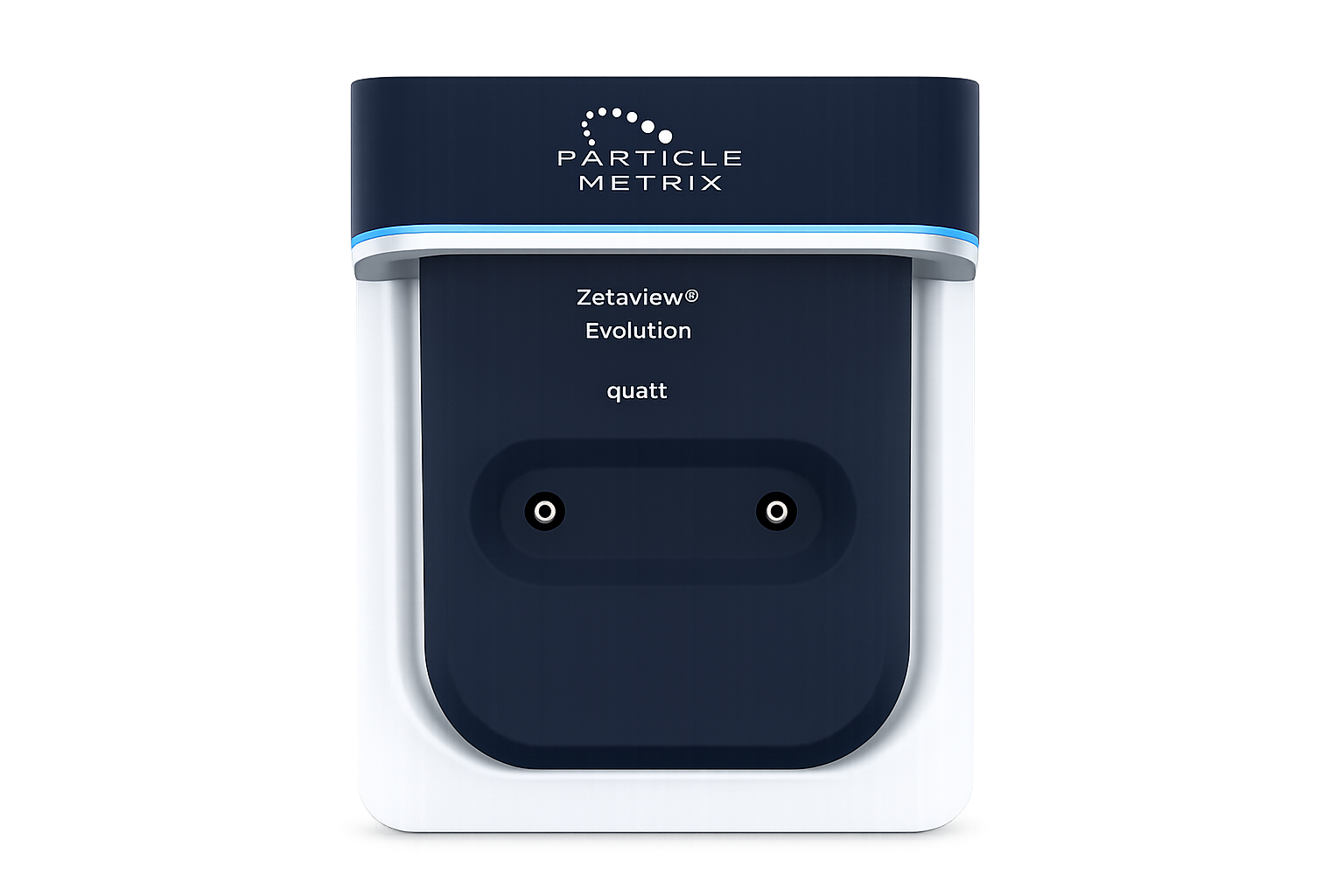
Next-Generation Particle Characterization Platform
Particle Metrix ZetaView evaluation platform integrates cutting-edge detection modules for complete biological particle analysis. Designed for reliability, reproducibility, and simplicity, it eliminates manual calibration and provides real-time quantitative data.

Next-Generation Digital Flow Cytometry Platform
Pangnostics delivers a breakthrough in particle analysis with its Digital Flow Cytometry platform-bringing single-molecule precision to complex biological systems. By overcoming the detection limits of conventional cytometry, it enables researchers to uncover hidden subpopulations and generate truly quantitative insights.
Two ways of seeing the nanoscale.
These platforms read biological particles through complementary physics — tracking how particles move, and characterizing the single molecules on their surface.
Nanoparticle Tracking Analysis
Up to four lasers illuminate the particles suspended in the cell, while a camera records the light each one scatters — and the fluorescence from labelled particles — as they move under Brownian motion. The Stokes–Einstein relation converts that motion into a hydrodynamic diameter, and counting the tracked particles in a known volume gives an absolute concentration — particle by particle, no calibration curve required.
Digital Flow Cytometry
Sample flows through a planar microfluidic channel where line-confocal optics and four lasers interrogate a tiny detection volume. Each fluorophore-tagged molecule is counted as it passes — across twelve channels — giving absolute, calibration-free numbers and per-particle phenotypes that bulk methods average away.
Most of the EV population hides below the floor.
Therapeutic and biofluid preparations are dominated by 30–70 nm vesicles. Where a method's detection floor sits decides whether you measure the real sample — or a brighter, biased subset of it.
Drag the detection floor — see the sample you'd actually measure
Illustrative model of a real EV population, where the smallest vesicles dominate. Single-molecule detection reaches ≤35 nm; raising the floor toward conventional limits hides the majority and inflates the apparent size.
| Method | Smallest particle | Label-free size | Per-particle | Absolute count | Copies / particle |
|---|---|---|---|---|---|
| Western blot / ELISA | bulk (no sizing) | — | ✕ | ✕ | ✕ |
| Conventional flow | ~500 nm | ✕ | ✓ | relative | ✕ |
| High-sensitivity flow | ~70–80 nm | ✕ | ✓ | bead-calibrated | semi |
| Nanoparticle tracking (NTA) | ~10 nm | ✓ | ✓ | ✓ | ✕ |
| Single-molecule dFC | ~ 1 nm | fluorescence | ✓ | ✓ calibration-free | ✓ |
* NTA sizes and counts label-free; single-molecule dFC adds fluorescence phenotyping and surface-marker copy number. Simplified for comparison — not a substitute for method validation.
From bulk averages to single molecules.
Each step in EV characterization recovers detail the previous one averaged away.
Bulk averaging
Western blot & ELISA confirm a marker is present — but only as a population average, blind to how it's distributed.
Particle tracking
NTA reads Brownian motion to give label-free size, concentration, and zeta — particle by particle.
Single-EV phenotyping
High-sensitivity flow resolves markers on individual vesicles — but a detection floor still hides the smallest EVs.
Single-molecule digital
Pangnostics dFC counts individual molecules to ≤35 nm — absolute, calibration-free, with copies-per-particle.
Small particles, outsized roles.
Extracellular vesicles ferry proteins and RNA between cells — making them biomarkers, drug-delivery vehicles, and therapeutics in their own right. The same analysis extends across the nanoscale of life.
Extracellular Vesicles
Size, concentration, and surface markers — with per-EV phenotyping.
Lipid Nanoparticles
LNP analysis for drug delivery and mRNA therapeutics.
Nanobubbles
Size, charge, and stability across diverse media.
Viruses & VLPs
Characterisation for diagnostics and vaccine development.
Environmental
Tracking nanoparticles for ecological impact studies.
Instruments and reagents, end to end.
Pangnostics dFC
Single-molecule digital flow cytometry — absolute quantitation beyond the detection limits of conventional cytometry.
Explore →ZetaView® Evolution
Multiparameter NTA with calibration-free concentration, size, and zeta in one workflow.
Explore →Lyophilized Exosomes
Stable, reconstitutable EVs for calibration and method development.
View →F-NTA Antibodies
Targeted tetraspanin detection antibodies — CD9, CD63, CD81 — for fluorescence NTA.
View →Method Qualification
Scientific, regulatory, and engineering support to bring a method to your biology.
Talk to us →Let's map the right measurement to your biology.
Start a conversationFor Research Use Only. Not for use in diagnostic procedures.